Medical imaging
A peek inside the body without opening it
Medical imaging is widely used in medicine. Yet it is a relatively new discipline in animal research. Medical imaging offers the possibility to peek inside the body without having to open it. For the animals this means less suffering. Non-invasive techniques also offer the possibility to follow the animals over time and fewer animals are needed for an experiment.
Medical imaging therefore is part of the 3Rs program and that is why BPRC has invested in these techniques in recent years. We explain these below.
X-ray
Soft tissue absorbs x-rays but bone tissue does not. On an X-ray picture, bones light up while the soft tissue remains dark. X-ray is indispensable for the diagnosis of for instance bone fractures. Veterinarians at BPRC use x-rays during health surveillance of the animals.
Ultrasound
Sound waves reflects differently on hard and soft tissue. This allows us to visualize tissue and organs. Ultrasound is often used to visualise pathological abnormalities, but also for prenatal examination. BPRC uses ultrasound for diagnostic purposes and health monitoring.
Ultrasound scan
Where normal ultrasound examination takes place on bare skin with guide gel, there is also the option to use ultrasound for internal examination, for example during intestinal diagnostics. Ultrasound scan, for example, supports experimental tuberculosis and influenza research.
PET-CT
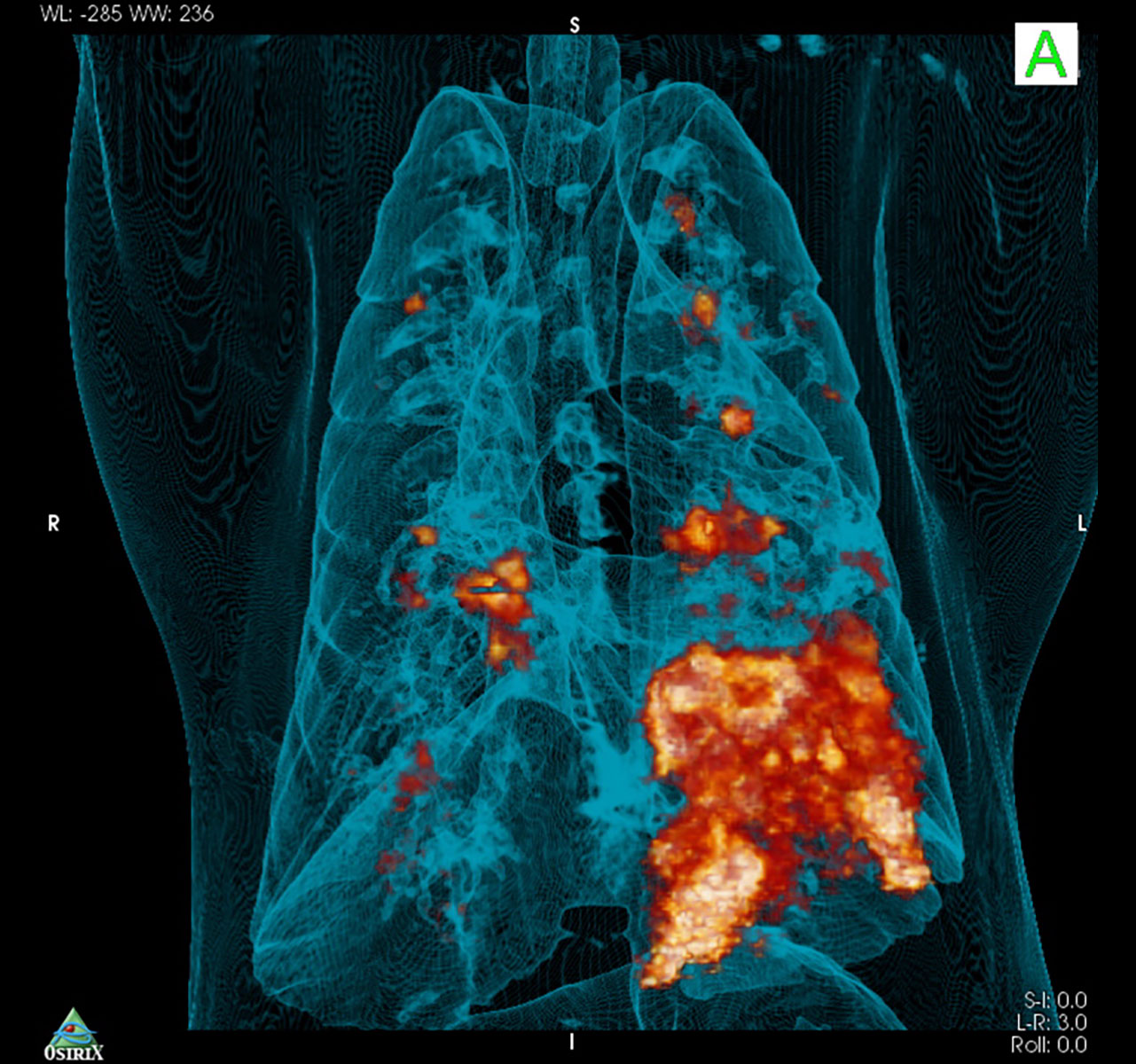
X-ray and ultrasound give anatomical information but do not say anything about the function. PET-CT does make that possible. The CT scanner uses the same X-rays as a 'normal' X-ray. But the CT scanner only makes cross sections of the body. All the photographed slices in succession form a three-dimensional image. Specific functions can be visualized with a radioactive contrast fluid. For example, immune cells use a lot of energy. By labelling the source of the energy, the glucose, with a radioactive substance, active areas of the immune system are visualised. PET-CT is therefore increasingly used to support experimental animal research.
FLIR
An infrared camera does not detect light but warmth. One of the hallmarks of inflammation is heat. With an infrared camera we can visualize local reactions, for example after vaccination.